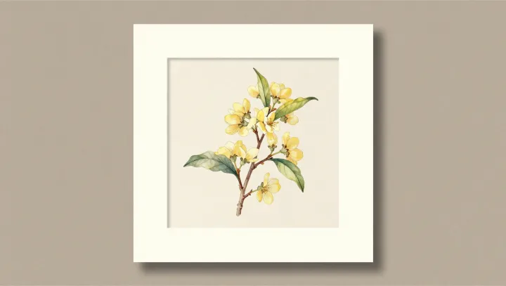
A painted twig should be unreliable evidence. Yet many eighteenth‑century botanical plates still let modern taxonomists pin a Latin name on a living plant with unsettling confidence. That accuracy rested on a quiet alliance between studio practice, early microscopy and the rise of formal plant anatomy.
The core trick was not beauty but redundancy. Artists working with botanists used simple lenses and compound microscopes to study venation, trichomes, anthers and ovules, then folded those microscopic traits back into a single branch or rosette. One sheet might show a flowering stem, a cross‑section of ovary, a magnified seed and a dissected corolla, each rendered at a different scale but locked to the same specimen through field notes and accession numbers.
What looks like ornament is often code. The exact angle of leaf attachment, the number of stamens, the pattern of secondary veins, even the contour of a seed scar were chosen to match emerging taxonomic keys, long before DNA barcoding or phylogenetic trees. Many illustrators worked directly from herbarium vouchers and repeated dissections, not from memory, so diagnostic morphology was copied again and again until it became almost schematic.
The result is a hybrid record. Part portrait, part anatomical chart, part data sheet. Modern botanists cross‑check these plates against preserved specimens and find the same combination of characters, which is why a centuries‑old painted branch can still stand in for a missing plant in a taxonomic argument.