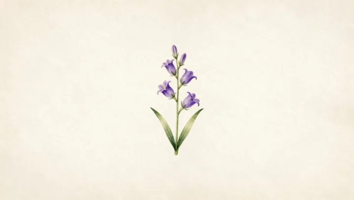
A lone purple bellflower on paper was often more forensic than its photographic contemporaries. Under early lenses and silver emulsions, petals washed out, faint venation vanished, and low‑contrast structures drowned in glare, while the watercolorist could boost those barely visible veins and hairs with saturated pigment and controlled line.
The real advantage sat in selective exaggeration. Where orthochromatic plates mis‑registered violet as near black and blurred depth of field, the artist could keep corolla lobes, anthers, and the stigma all in notional sharp focus, rendering each organ with quasi‑diagrammatic clarity that no single exposure and aperture combination could achieve under field conditions.
Ecology also hid in those painted margins. Photographs clipped by slow shutter speeds and rigid framing often lost the neighboring moss, soil texture, and companion plants, yet the illustrator could annotate habitat by choice: a specific leaf litter color, a precisely angled stem showing phototropism, or insect damage on sepals that hinted at herbivory and seasonal pressure on the species.
What looks like art history is in fact an imaging trade‑off. Early optics, emulsion spectral sensitivity, and low dynamic range imposed hard constraints, while brush, graphite, and human visual cognition formed an analog, loss‑correcting pipeline that stored both morphology and micro‑habitat in one deliberately edited bellflower study.